Magnetic resonance imaging (MRI) is a sophisticated test healthcare providers use to help diagnose certain medical conditions and injuries. It involves taking detailed photos of the inside of your body.
“It’s a cross-sectional imaging technique that uses a combination of magnetic fields and radio frequency fields to generate an image,” says Bruce Railey, MD, a radiologist with Medical City Arlington in Arlington, Texas. One MRI test can produce dozens of images—each is called a slice—of the body part being studied.
Because having an MRI involves sliding your body into the machine, the procedure can be intimidating for some people.
But there’s no reason to worry. Getting an MRI is a painless process that takes less than an hour in many cases, according to Dr. Railey.
Who needs an MRI?
Your healthcare provider (HCP) may send you for an MRI for a variety of reasons. It’s mostly used to look at soft tissue like muscles and organs, as opposed to bones, according to Railey.
“One of the MRI’s original applications was imaging of the brain and spinal cord, and that is still a very common use,” he says. “But it has also become very valuable in imaging of tendons, ligaments, joint cartilage, and other soft tissue. It’s become instrumental in imaging of the liver because it’s very sensitive in detecting if a tumor has spread from the liver to other sites.”
Know before you go
Insurance companies may require a preliminary test, such as an X-ray, before they will cover an MRI. “That’s not uncommon with insurance companies, but it’s not what a physician would consider a requirement,” says Railey. “An X-ray may show an abnormality of a bone, but the MRI is far more sensitive in detecting anything with the muscles, tendons, ligaments, or cartilage.”
You may need to stop eating and drinking four to six hours before the imaging, depending on what body part is being scanned. For some procedures, especially tumor imaging, you may need a contrast dye administered intravenously (through an IV). “The dye can increase the contrast between normal and abnormal areas of tissue,” Railey says.
While there’s no evidence that an MRI would harm a pregnant woman or a fetus, the use of a contrast agent called gadolinium should be limited, according to the American College of Obstetricians and Gynecologists. If you are slated to get an MRI, tell your HCP if you’re pregnant, or if you have a metallic implant such as a pacemaker or an infusion pump inside your body.
What to expect during the procedure
The procedure takes between a half hour and an hour, depending on how many parts of your body are being scanned. You may need to wear a hospital gown if your clothes have metal pieces on them, such as zippers or buttons. You’ll also need to remove any metal jewelry. Imaging facilities typically provide lockers for storing valuables.
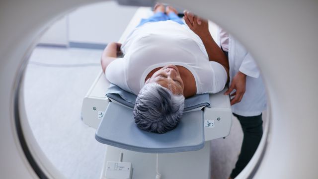
Most MRI machines have a tunnel in the middle and are open on both ends. You’ll lie on your back on a table that slides inside the tunnel. It’s a tight squeeze and may cause claustrophobia in some people. Some facilities have MRI machines called open or standup MRIs, which allow you to sit, stand, or bend in a less-confined area. They can be helpful for patients with claustrophobia, obesity, or movement issues.
Once you’re ready for your MRI, the technician will position the body part to be scanned. It’s important to stay still throughout the procedure so the images don’t come out blurry. The machine intermittently makes a lot of noise, so you will likely be given earplugs or headphones with music.
“Most people are okay, but the noise and claustrophobia are the two most common complaints,” says Railey. There will be a technician in the room running the machine, and you will be able to communicate with them via a two-way radio, Railey says.
Tell your HCP if you are prone to claustrophobia; taking a mild sedative before the test is not uncommon. If you take medication to be sedated, you won’t be able to drive yourself home. Bring a family member to the testing center if you think you’ll need medication.
After the test
Once the study is completed, the machine generates the images. “It then becomes the radiologist’s job to look at the images in conjunction with the patient history and the reason for the study,” says Railey. The radiologist then creates a written report, which is sent to the provider who ordered the MRI. Your HCP will discuss the results with you, along with any next steps that may be required.


