Updated on April 30, 2024.
In the United States, more than one-third of women 40 and older don’t get their routine mammogram, according to the Centers for Disease Control and Prevention (CDC). Supporting breast cancer awareness and prevention efforts, such as wearing pink ribbons and donating to cancer charities, can help create awareness and fund critical research. But routine screening is another important step millions need to take to help increase early detection of the disease.
Here are some answers to some of your top questions about getting mammograms, from making your appointments to getting results and why it’s important to get the test done at recommended intervals or as discussed with your healthcare provider (HCP).
Why do people miss or delay mammograms?
Some people miss or delay getting their recommended mammogram for various reasons. These might include factors such as language and cultural differences, concern about out-of-pocket costs, fear mammograms will be painful or expose them to radiation, and apprehension their test results will say they have cancer. Some are embarrassed to get a mammogram while others don’t trust the healthcare system.
This limited access to care exacerbated health inequities and disparities in screening mammography among people with low incomes and from certain racial and ethnic minority groups. This includes people from African American and Hispanic backgrounds.
Some don’t have HCPs they usually go to and haven’t been referred for a mammogram. Others have been misinformed about the benefits and risks of mammograms or don’t know about breast cancer risks or when and how to screen for the disease.
Other barriers to getting a mammogram might include a lack of:
- Childcare or care for an adult or parent who depends on you
- Sick leave or time off work
- Transportation to and from the imaging center
Why get a recommended mammogram?
Breast cancer is the most commonly diagnosed cancer among women in the U.S., second only to skin cancer that isn’t melanoma. It’s the second leading cause of death due to cancer among U.S. women overall, trailing only lung cancer. Among Black and Hispanic women, it tops the list as the leading cause of cancer death.
Mammography is a primary screening tool for detecting breast cancer. In fact, the disease is highly curable when caught early.
Getting a mammogram at recommended intervals is one of the most effective ways to detect breast cancer early. For some, the test finds breast cancer up to three years before it can be felt, notes the CDC.
When and how often to get a mammogram?
Various professional health and medical organizations publish recommendations for screening mammograms. For instance, the U.S. Preventive Services Task Force (USPSTF) recommends women aged 40 to 74 years get a mammogram every other year.
The American Cancer Society (ACS) recommends women age:
- 40 to 44 be given the option to start screening with a mammogram each year
- 45 to 54 get their mammogram yearly
- 55 and older be given the choice to continue with their annual mammograms or switch to getting them every other year
These recommendations apply to women with an average risk of breast cancer. In general, you’re at average risk if you haven’t had chest radiation therapy before 30 years old and don’t have a:
- Personal history of breast cancer
- Strong family history of breast cancer among close biological relatives, such as your siblings and parent
- Gene mutation known to raise the risk of breast cancer, such as in breast cancer (BRCA) genes 1 or 2
It’s important to talk with your HCP about your risk factors. They can provide more details about these factors and whether you’re low, average, or high risk for breast cancer. They can also discuss when to start and how often to get a routine mammogram.
Are mammograms recommended after 74 years old?
Some professional organizations differ in their stance when it comes to getting a mammogram past 74 years old. Some, such as the USPSTF and American Academy of Family Physicians, don’t provide a recommendation for or against continued screening after this age, citing a lack of evidence either way.
But the ACS and American College of Obstetricians and Gynecologists specify that screening may continue as long as you’re in good health overall and have a life expectancy of at least 10 years.
Despite these varying recommendations, all U.S. professional guidelines support a tailored approach to deciding whether screening should continue beyond age 74. This includes having a frank talk with your HCP about the benefits and harms of continued screening and your individual risk factors and preferences, and deciding together which approach best suits your needs.
When is the best time to schedule a mammogram?
Book an appointment for when your breasts are less likely to be sore or swollen. You may feel less discomfort during the scan, and it can help your technician get better images.
If you haven’t reached menopause, the week after your period is generally a good time. Your breasts may feel more sensitive the week before you menstruate and during your period. Therefore, this timeframe may not be the best time to get it done.
While scheduling your appointment, let the imaging center know if you have any physical limitations or disabilities that must be taken into account. These include issues such as not being able to raise your arms or breathing problems like asthma.
The staff can make plans to accommodate your needs or refer you to another facility better equipped to manage your care.
Will my insurance cover my mammogram?
Thanks to the Affordable Care Act (ACA), signed into law in 2010, most women between the ages of 40 and 74 are covered for screening mammograms every one to two years, if they have private health insurance or additional insurance coverage from states with Medicaid expansion programs.
Medicare will cover one baseline mammogram between the ages of 35 and 39, and then an annual screening mammogram every year after that. There’s no cost for screening mammograms if your HCP orders them.
For people without insurance, low- or no-cost screenings may be available through reputable health organizations like the Susan G. Komen Foundation and the National Breast Cancer Foundation.
Coverage for diagnostic mammograms varies by plan. These X-rays are performed after your HCP detects a lump in your breast or you have symptoms that may signal breast cancer.
Diagnostic mammograms may involve a co-pay, or you may have to meet your annual deductible first. Before making an appointment, reach out to your insurance company for details. Medicare will cover 80 percent of the cost of “medically necessary” diagnostic mammograms after you meet the Part B deductible.
What are the advantages of 3D mammograms?
Digital breast tomosynthesis (DBT), or 3D mammograms, are more advanced than conventional 2D mammograms. With conventional mammograms, the volume of each breast is compressed into a single 2D image. DBT divides the volume of each breast into multiple segmented images called slices.
Viewing these thin slices together creates 3D images of your breasts. DBT can be used alone or alongside other types of mammograms.
You’re less likely to need additional imaging tests after getting DBT since it provides a more detailed view of your breasts. The 3D images make it easier to see various changes to your breasts, especially if you have dense breasts.
Having dense breasts means you have less fatty tissue and more glandular tissue meant to produce and excrete breast milk, as well as more fibrous or supportive tissue called stroma surrounding these glands. About 50 percent of women aged 40 and older have dense breasts, which increases their risk for developing breast cancer. Getting a mammogram is the only way to confirm whether you have dense breasts.
But 2D mammograms can’t distinguish between cancer and dense breast tissue since both appear white on these X-rays. 3D mammograms can see beyond these dense areas to detect cancer more accurately while reducing the likelihood of false-positive results.
And because DBT’s more sensitive, they’re more likely to find lower grade cancers. These cancers grow more slowly and are less likely to spread to other parts of your body than higher grade ones.
Medicare will pay for 3D mammograms as long as they’re performed at the same time as 2D mammograms. If you don’t have Medicare, check with your insurance provider regarding 3D mammograms, however, as some plans may not pay for them.
How to prepare on the day of the mammogram?
Wait to apply deodorant or antiperspirant to your upper body area until after your mammogram, as this may interfere with the imaging. Skip the lotion, cream, oil, powder, and perfume around your breasts and upper arms, as well. These may produce white spots on the images and can make it harder for the mammogram to compress your breasts.
Leave necklaces at home and consider wearing a two-piece outfit so you can remove your top for the imaging while keeping clothes on the lower half of your body. Instead of a one-piece dress or jumpsuit, opt for a shirt with pants, shorts, or a skirt.
What can I expect during my mammogram appointment?
Once you arrive for your appointment and before the actual imaging test starts, you’ll likely answer background questions related to your:
- Personal and family medical history
- Previous medical or surgical procedures
- Recent or current medications—including over-the-counter (OTC) and prescription medicines, essential oils, vitamins, and other herbs and dietary supplements
If you’ve discovered any changes in your breasts since your last mammogram, be sure to let your HCP and mammogram technician know. The same thing goes if you’re pregnant, breastfeeding, or have breast implants.
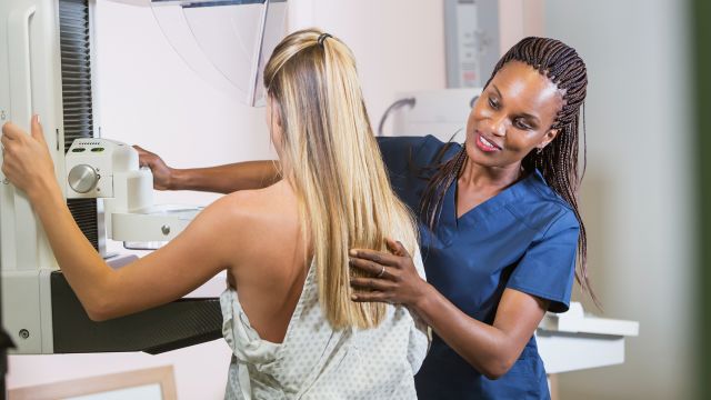
Once you’re in the exam room where the mammogram will be performed, your technician will have you remove clothes covering your upper body. They may also use markers on your skin to identify the position and location of your nipples, moles, masses and other prominent skin features or areas specified by the HCP who ordered your mammogram.
One of your breasts will then be placed on a small platform attached to the mammogram machine, where it will be slowly compressed between this lower platform and an upper plate. You’ll be asked to stay still and hold your breath while the technician takes images.
Once this first image batch is complete, the technician will release the upper plate and reposition your breast to obtain images from a different angle. The entire process will be repeated for your other breast.
If it’s a diagnostic mammogram, they’ll focus largely on the areas of breast tissue in question.
Are mammograms safe while pregnant or breastfeeding?
In general, mammograms are considered safe during pregnancy and while breastfeeding. This is supported by current guidelines on breast imaging of pregnant and lactating women published by the American College of Radiology (ACR). In fact, the ACR notes DBT may be beneficial for women who are pregnant or breastfeeding as it’s more likely to spot cancer through dense breast tissue.
The X-ray uses small amounts of radiation, which target the breasts and lessens the chances of radiation exposure to other areas such as your womb. During the procedure, your mammogram technician will cover your lower belly with a lead shield for added protection.
Despite the small amount of radiation, your HCP can’t say with full certainty what the effects may be on your fetus. It’s best to discuss the risks and benefits of the imaging test with your HCP in advance and come to a decision together.
Do mammograms hurt?
For some people, mammograms can cause discomfort. Your breasts are spread and squeezed to get a complete picture of your breast tissue. This may be uncomfortable. But it’s quick.
Your breast is compressed for about 10 to 15 seconds for each image. If you’re concerned or hesitant, speak with your HCP about whether you can take an over-the-counter pain reliever, such as ibuprofen, an hour or so before the mammogram.
And during the mammogram, let your technician know if the pain is too much to tolerate. They can take a short break from the procedure or adjust the device as needed.
How long will the mammogram take?
All told, the process of getting a mammogram typically lasts 10 to 30 minutes. Certain factors can affect how long the procedure takes. These include breast:
- Density
- Implants
- Masses and other suspicious growths
- Size
When will I get my mammogram results?
Once you’ve completed your mammogram, a radiologist will review and interpret your images and send a detailed report of their findings to your HCP. These medical doctors are trained to diagnose injuries and disease using medical imaging devices such as X-rays, magnetic resonance imaging (MRI), ultrasounds, and other radiology techniques.
In the United States, your HCP is legally required to provide you with a written summary of your mammogram results within 30 days of the procedure. Most of the time, you’ll get results much faster, though.
You’ll usually get your results back within one week of the test. If your radiologist finds something to suggest the presence of cancer, they must share results with your HCP as soon as possible. Be sure to reach out to your HCP or imaging center if you don’t get your results back after 10 to 14 days.
As of March 2023, the U.S. Food and Drug Administration (FDA) requires mammography facilities to also notify patients about the density of their breasts.
What are the next steps after getting my mammogram?
If your mammogram is normal, continue to be screened as discussed with your HCP. Between screenings, speak with your HCP if you notice any changes affecting the look, feel, or shape of your breasts.
If cancer is suspected based on your mammogram results, your HCP will order more tests to confirm the diagnosis or rule it out and determine another cause. This often involves a more detailed look at the area in question with a diagnostic mammogram.
Your HCP may also order one or more of the following tests:
- Breast biopsy to confirm or rule out breast cancer conclusively. This involves removing a small piece of breast tissue from the area in question and assessing it under a microscope.
- MRI, which can provide more accurate images for people with a family history of breast cancer, dense breast tissue, or other risk factors. This involves lying in a large tube-like imaging scanner while images are taken of your body’s organs, tissues, and internal structures with the help of a large magnet and radio waves.
- Ultrasound, which can distinguish between breast cysts and solid or calcified masses. This involves placement of a device called a transducer or probe on the surface of your skin to create live images or videos of your internal organs and soft tissues with the help of high-frequency sound waves.
Following up with additional tests can help provide greater clarity. And if it is cancer, mammograms can help find breast tumors at an earlier stage, when they are often easier to treat.


